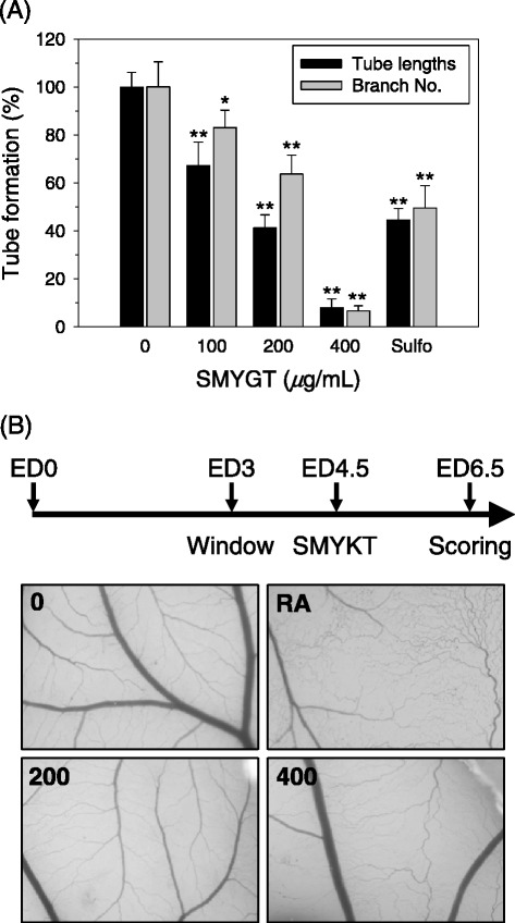
Figure 1.

Anti-angiogenic potential of SMYGT in vitro and in ovo. (A) HUVECs cultured on BME-pre-coated supports were exposed to different concentrations of SMYGT for 12 h. The tube length and branch numbers were determined by analyzing digitally captured images. Sulforaphane (Sulfo, 5 μM) was administered in parallel as a positive control drug. Relative tube formation was determined by comparing each group with the vehicle treated group (0 μg/mL SMYGT), and the data are presented as the means ± standard deviation (S.D.) of triplicate experiments. *P < 0.05, **P < 0.01, ***P < 0.001. (B) At ED4.5, A quarter size of thermanox coverslips containing variable amounts of SMYGT (0, 200, 400 μg/disc) were applied to the CAMs. After 2 days of incubation, 10% skimmed milk solution was injected into the CAM for observation of the inhibition zone of angiogenesis and digital images were captured. Retinoic acid (RA, 1 μg/disc) used as a control for inhibition of new vessel formation.
