
Figure 2.
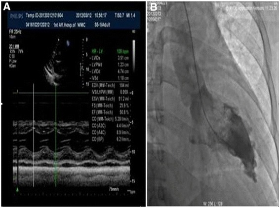
Echocardiography and left ventricular angiography. A. Echocardiography revealed normal movement of the ventricular wall and deteriorated diastolic function (left ventricular end diastolic diameter (LVEDD), 47 mm; interventricular septum thickness (IVS), 12 mm; left ventricular ejection fraction (LVEF), 50.8%). Coronary angiography showed that there was no stenosis or obstructive lesions in the coronary artery; B. Left ventricular angiography showed thickening of the apical ventricular membrane.