
Figure 5.
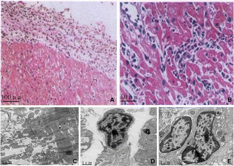
Endomyocardial biopsy and cardiac electrogram. Endomyocardial biopsy (A and B) showed there was obvious infiltration of EC and focal necrosis. Electron microscopy (C, D and E) revealed extensive myocardial interstitial infiltration by EC accompanied by focal myocardial fiber cracking and disintegration Internal scales: A: 100 μm; B: 50 μm; C, D and E: 1 μm.