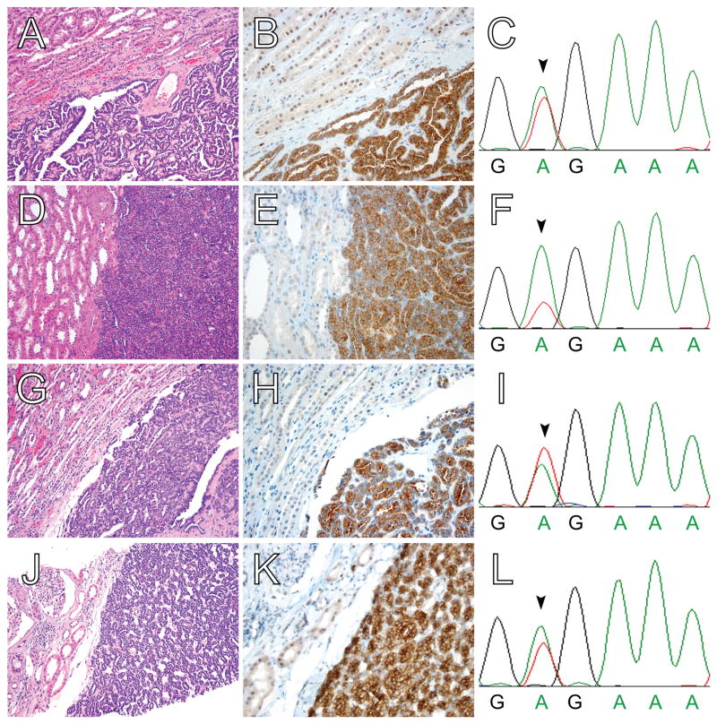
Figure 1. BRAF V600E protein expression in MA, as detected by IHC with a mutation-specific antibody, is highly correlated with the presence of BRAF V600E mutation by Sanger sequencing.
(A,D,G,J) H&E, (B,E,H,K) BRAF V600E IHC, and (C,F,I,L) BRAF exon 15 genomic sequencing of four MA cases. All four cases show the typical histomorphologic features of MA and demonstrate diffuse, moderate to strong cytoplasmic BRAF V600E staining. Adjacent non-neoplastic kidney parenchyma shows negative to weak cytoplasmic staining and weak to moderate nuclear staining. In each case, Sanger sequencing of BRAF exon 15 reveals a corresponding thymidine to adenine substitution at codon 600 (arrowheads in C,F,I,L); this substitution results in the missense BRAF V600E mutation. Sanger sequencing confirmed the presence of BRAF V600E mutations in the remaining three MA cases with diffuse, moderate to strong cytoplasmic BRAF V600E staining (data not shown). No other BRAF exon 15 mutations were identified in these seven cases. Magnification: (A,D,G,J) 20X; and, (B,E,H,K) 40X.

