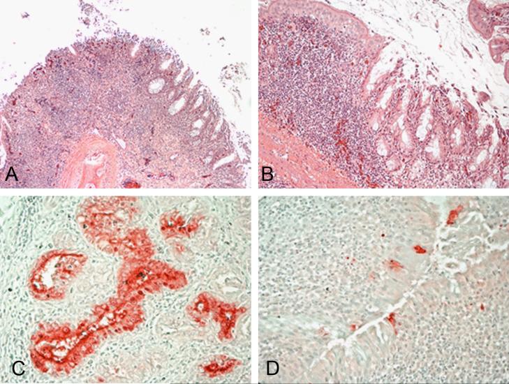
Figure 3.
Histopathology and immunohistochemical staining for avian influenza virus antigen in tissues of chickens infected by the ICh route with the A/Anhui/1/2013 H7N9 virus, 3 and 6 DPI. Photomicrographs, magnification 200-400X; virus staining in red. Nasal turbinates. Severe necrotizing rhinitis with submucosal congestion and edema, glandular hyperplasia, and lymphoplasmacytic infiltration (A and B). Demonstration of viral antigen in the epithelial cells of nasal glands (C) and nasal epithelium of the turbinates (D).

