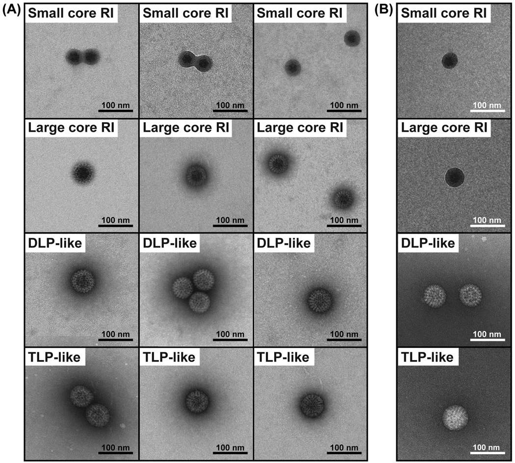
Fig. 5. EM imaging of particles in gel-purified core RI population.
(A) Three independent EM images each of four general particle types seen in the gel-purified core RI population. The grids were prepared with a thin layer of metal stain. Scale bar is 100 nm. (B) A representative EM image of each general particle type from grids prepared with a thick layer of stain. Scale bar is 100 nm.

