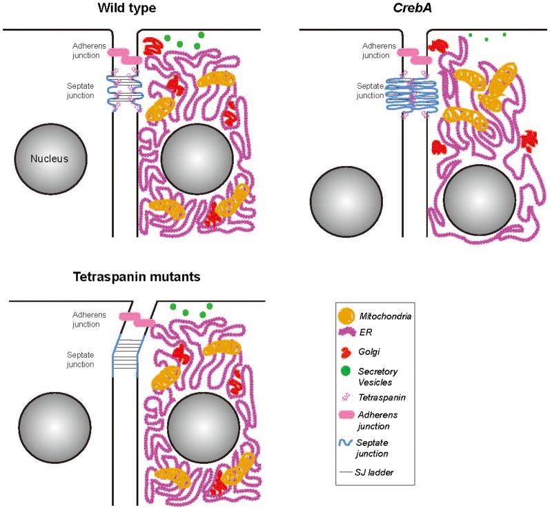
Fig. 8. Cartoon representation of WT, CrebA and tetraspanin mutant cells with the differences in organelle localization and SJ structure highlighted.
In WT SG epithelial cells, the SJs are slightly convoluted and there is an accumulation of tetraspanin proteins specifically at the SJ region. In CrebA mutants, there is a global reorganization of membranous organelles and the SJs become more convoluted, due to excess membrane. By microarray analysis, there is an increase in tetraspanin gene transcription presumably resulting in more tetraspanin protein at the SJ to stabilize the increased SJ membrane folds. In tetraspanin deficient cells, the SJs are slightly longer and less rigid, resulting in a swaying phenotype.

