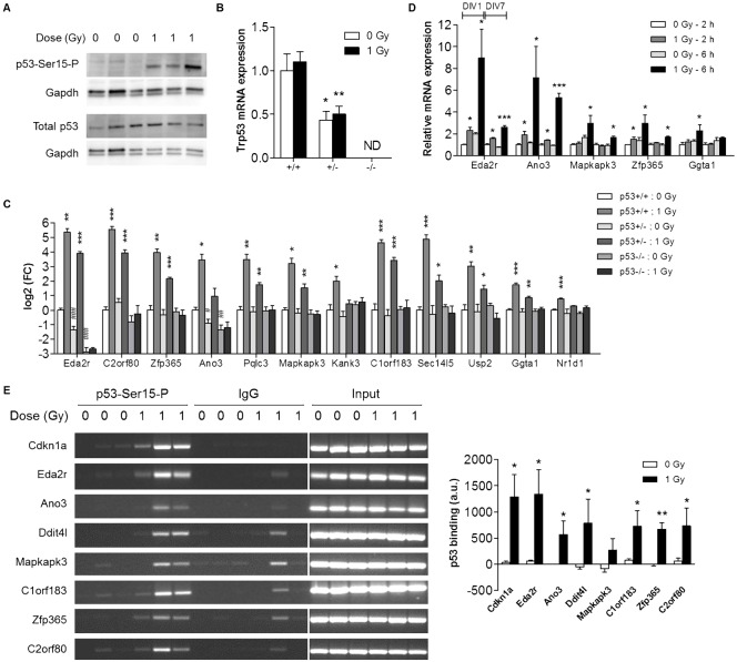
Fig. 4. p53-dependent expression of novel target genes.
(A) Western blotting was performed using antibodies against the phosphorylated (p53-Ser15-P) as well as the total form of p53. (B,C) mRNA expression was determined by qRT-PCR in E11 brains from p53+/+, p53+/− and p53−/− littermates (n = 4) at 2 h post-irradiation. Please note the logarithmic scale of the Y-axis in B. *p<0.05; **p<0.01; ***p<0.001 for the difference between control and irradiated mice from the same genotype (Student's t-test). #p<0.05; ##p<0.01; ###p<0.001 for difference with control p53+/+ mice (Student's t-test). (D) mRNA expression was determined by qRT-PCR in primary cortical neuron cultures (1 DIV and 7 DIV) at 2 h and 6 h post-irradiation (n = 3–4). *p<0.05; ***p<0.001 (Student's t-test). (E) ChIP-PCR was performed on pooled brains from individual litters (n = 3) of in utero irradiated mice at 2 h post-irradiation. Densitometric analysis of gel electrophoresis bands was performed using ImageJ. *p<0.05; **p<0.01 (paired Student's t-test). In all panels data indicate mean+s.e.m. ND, not detected; DIV, days in vitro.

