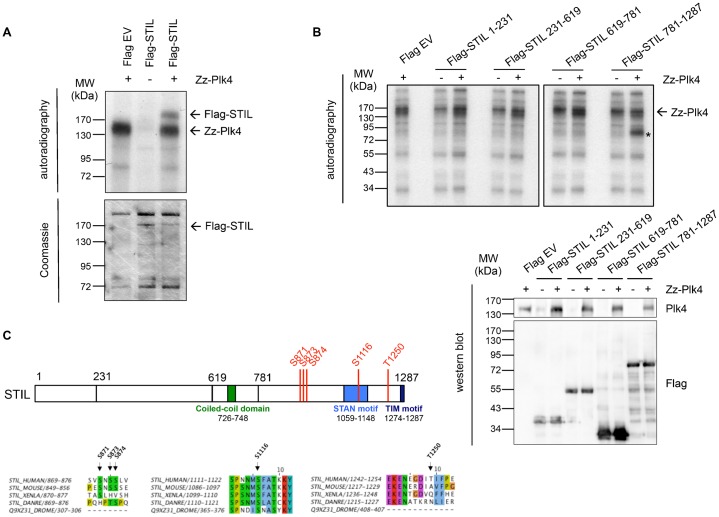
Fig. 6. Phosphorylation of STIL by Plk4.
(A) Full-length Flag-STIL expressed in HEK293T cells and immunoprecipitated with anti-Flag antibodies was incubated with bacterially expressed Zz-Plk4 in the presence of [γ-32P]-ATP. In vitro kinase assay with Flag-STIL or Plk4 alone served as a control. Kinase assays were analyzed by SDS-PAGE, Coomassie Blue staining and autoradiography. (B) Indicated Flag-STIL fragments were expressed in HEK293T cells and immunoprecipitated with anti-Flag antibodies. Immunoprecipitation fractions were incubated with bacterially expressed Zz-Plk4 in the presence of [γ-32P]-ATP, followed by SDS-PAGE and autoradiography. In vitro kinase assay with Flag-STIL fragments or Plk4 alone is shown as control. The asterisk indicates phosphorylated Flag-STIL 781-1287. 10% of each precipitation fraction was analyzed by western blotting using anti-Plk4 and anti-Flag antibodies. (C) Plk4 phosphorylation sites in the STIL protein identified by mass spectrometry analysis of bacterially purified GST-STIL 1-619 and 619-1287 phosphorylated in vitro by Zz-Plk4. Alignment of the identified sites in human, mouse, Xenopus and zebrafish STIL and Drosophila Ana2 is shown.

