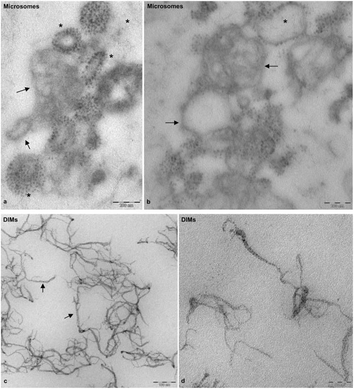
Fig. 3. Transmission electron microscopy of fixed, embedded and sectioned microsomes and DIMs.
(a,b) Sections of P2 membranes showing vesicles delimited by fainter membranes (arrows), seldom decorated with electron-dense particles (asterisks). (c,d) DIM membranes appearing as sharp, ribbon-like structures (arrows) with electrondense inclusions. Scale bars: 200 nm (a,b); 100 nm (c); 50 nm (d).

