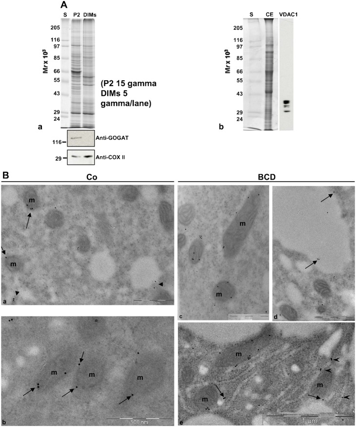
Fig. 6. DIM characterisation.
(A) Contamination of DIMs by plastid and mitochondrial membranes. (a) Anti-plastid (GOGAT) and anti-mitochondria (COXII) markers were probed on P2 (15 µg) and DIMs (6 µg). (b) The specificity of the anti-VDAC antibody probed on pollen tube crude extract. The antibody recognised four VDAC bands, identified by MS spectrometry. (B) Immunolocalisation of VDACs in pollen tubes. (a,b) In control pollen tubes the anti-VDAC antibody labelled the mitochondrial (mitochondria are indicated as m) outer membrane (arrows) and cytoplasmic vesicles (a, arrowheads). (c–e) In BCD-treated pollen tubes the anti-VDAC antibody still stained the mitochondrial outer membrane and vesicles (e, arrows), but also vacuoles (d, arrows) and ER (e, arrowheads).

