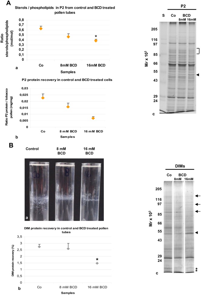
Fig. 7. Effect of BCD on DIM isolation.
(A) Effect of BCD on microsomal fraction. (a) Lipid analysis revealed a decrease in sterol content with 8 mM and 16 mM BCD treatment, with respect to control cells. (b) Less protein was recovered in P2 when pollen tubes were incubated with 8 mM and significantly less after incubation with 16 mM BCD with respect to control. (c) Electrophoretic analysis showed changes in the polypeptide pattern of 16 mM BCD-treated pollen tubes P2 (square bracket indicates decreasing polypeptides, arrowhead indicates enhanced polypeptide), with respect to control. (B) Effect of BCD on DIMs. (a) The amount of material in the floating band decreased when pollen tubes were incubated with BCD. The behaviour of DIMs also changed from fine particles in control cells to lamellae in 16 mM BCD-treated samples. (b) A significant decrease in DIM protein recovery was detected in 16 mM BCD-treated pollen tubes. Error bars indicate standard errors. (c) Electrophoretic analysis revealed changes in the electrophoretic pattern in DIMs isolated from 16 mM BCD treated pollen tubes (arrows and asterisks indicate decreasing polypeptides, the arrowhead indicates the increased polypeptide). Control, 8 mM and 16 mM DIMs were run in the same gel, but in non-adjacent lanes.

