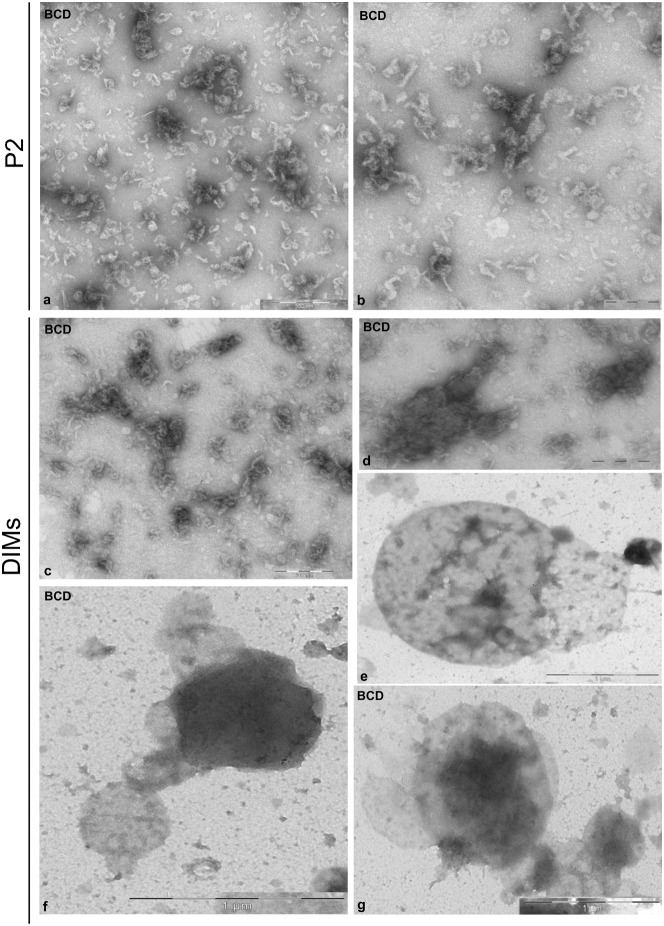
Fig. 8. Effect of BCD on P2 and DIM ultrastructure.
(a,b) The microsomal fraction appeared to be made up of membrane fragments, instead of organelles. (c–g) Isolated DIMs showed as clusters of membrane fragments (c,d) or as smooth vesicles with diameters of 0.5–1 µm (e–g). Scale bars: 200 nm (b-d); 500 nm (a); 1 µm (g,f).

