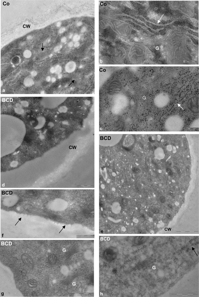
Fig. 11. Effect of BCD on pollen tube morphology.

(a–c) The cytoplasm of control pollen tubes was rich organelles such as mitochondria, Golgi bodies (a, arrow; b,c, indicated as G) and smooth (b, arrow) and rough ER (c, arrows). The cell wall (a, indicated as CW) showed fibrillar and amorphous components. (d–h) BCD-treated pollen tubes showed a strong reduction in cell wall fibrillar component (d,e). Alterations were observed in PM ultrastructure (f, arrows). Golgi bodies (g) and ER elements (h, arrow) were rarely observed in BCD-treated pollen tubes. Scale bars: 200 nm (b,c); 500 nm (a,d; f-h); 1 µm (e).
