
Figure 2.
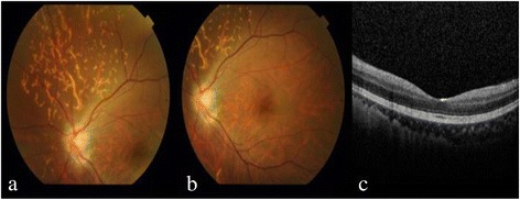
Fundus examination revealed optic disc edema and some linear whitish opacities over the superior and temporal sites in the left eye, suggesting multiple CaHA emboli in the choroid vessels (a). No macular edema in left eye was revealed on fundus examination (b), or OCT (c).