Figure 3.

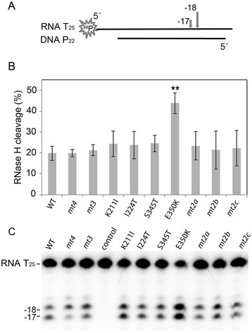
Qualitative RNase H assay. (A) Schematic representation of the 5′ 32P-RNA25/DNA22 hybrid. The arrows on top of the RNA indicate the major RNase H cleavage sites at position −17 and −18. The first nucleotide of the RNA hybridized to the 3′-OH nucleotide of the DNA strand is designated −1. (B) Quantification of the RNase H cleavage products. The diagram depicts the mean values and standard deviations (black bars) of three independent experiments. For quantification of the cleavage products the total amount of labeled RNA per lane was set to 100%. Only the p-value of E350K ≤ 0.01 (**) represents a statistically significant difference to the WT protein. (C) Autoradiogram of a typical RNase H cleavage experiment. RNase H reactions were performed with 240 nM 5′ 32P-RNA25/DNA22 hybrid and 50 nM RT-PR for 2 min at 25°C. RNA T25, uncleaved RNA; −17, 18 indicate the cleavage sites; control, reaction mix without enzyme.
