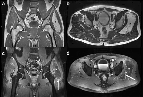
Figure 1.

48-year-old man with hibernoma of the left upper leg. Tumour between the gluteus medius and minimus muscles (star in image 1 a-d). MRI shows an isointense tumour compared to fat with slight rim enhancement (arrow in c) and a prominent vessel within the mass (arrow in d). a plain T1-weighted-TSE (TR 819 ms/ TE 11 ms), b plain T2-weighted-TSE (TR 5050 ms/ TE 96 ms), c contrast enhanced fat saturated T1-weighted-TSE (TR 895 ms/ TE 11 ms), d contrast enhanced fat saturated T1-weighted-TSE (TR 680 ms/ TE 11 ms).
