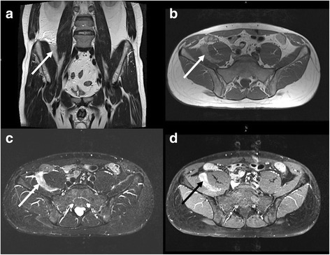
Figure 4.

MRI of the same patient as shown in Figure 3 . The tumour (arrows) is slightly hypointense compared to mesenteric fat in T2- (a, TSE, TR 5040 ms/ TE 137 ms) and T1-weighted images (b, TSE, TR 651 ms/ TE 11 ms). The T2-weighted TIRM sequence (c) shows marked hyperintensity of the mass (c, TR 5000 ms/ TE 74 ms/ TI 170 ms). After administration of contrast medium heterogeneous enhancement occurs (d, fat saturated TSE, TR 600 ms/ TE 11 ms).
