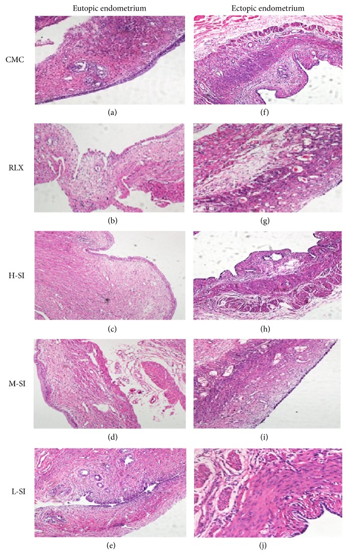
Figure 8.
Pathological morphology of endometrium and ectopic endometrium tissues of five-group rats after drug intervention analyzed with light microscopic analysis. As is shown above, in M-SI group, the number of gland and stromal cells of ectopic endometrium was decreased and the shape of the endometrium was nearly natural which were all meaningful differences compared with the other groups.

