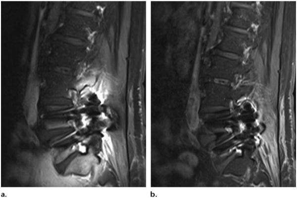
Figure 15.
Sagittal T1-weighted MR image with chemical fat saturation (TR, 510 msec; TE, 10 msec) (a) and Dixon (Siemens) MR image (TR, 692 msec; TE, 12 msec) (b) of the lumbar spine obtained in a patient with metal artifacts show that susceptibility artifacts are significantly greater with CHESS than fat-water separation. (Images courtesy of Rory Johnson and Peter Cazares.)

