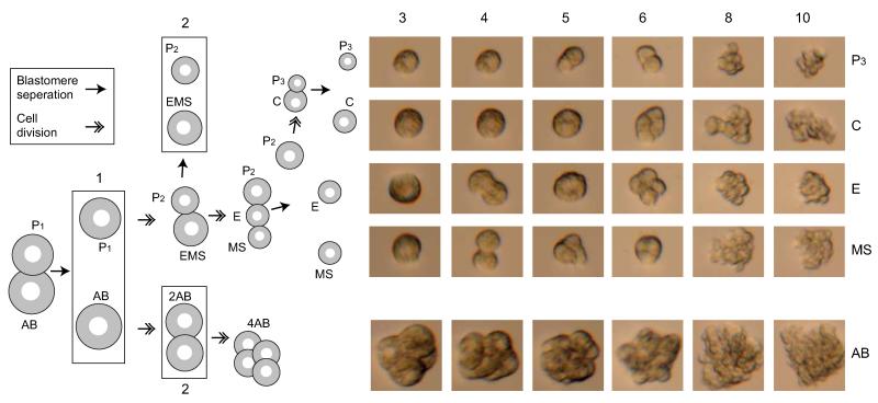
Extended Data Figure 1. In vitro culturing of the C. elegans embryonic founder blastomeres.
The cells are separated as shown in the left schematic and then cultured in embryonic growth medium11 as shown in the micrographs on the right. The numbers indicate the stages in which the cells were collected for transcriptome analysis. Six of the eleven stages are shown in the micrographs.

