Fig. 1.
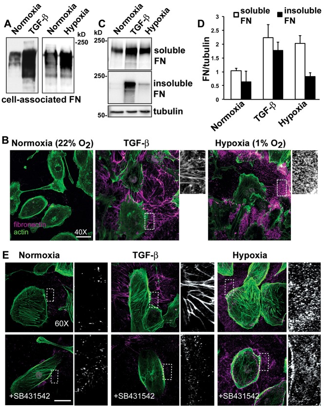
Hypoxia increases fibronectin abundance but only induces limited fibrillar assembly by HK2 cells. HK2 cells were untreated (normoxia controls), treated with TGF-β (10 ng/ml) in a normoxic environment or maintained at 1% oxygen (hypoxia) and for 48 hours. (A) Immunoblots of cells lysates for fibronectin (FN) under the indicated conditions. (B) Extracellular fibronectin was labeled before cell permeabilization (magenta) and actin filaments were labeled with phalloidin (green) after permeabilization. (C) Immunoblot of cell lysates separated into DOC-soluble and insoluble fractions. Immunoblotting for α-tubulin was used as a loading control. (D) Quantified fibronectin immunoblot signal expressed relative to α-tubulin loading control shown as mean±s.e.m. of three determinations. (E) Immunolabeling, performed as described in B, for extracelluar fibronectin for the indicated conditions in the absence and presence of the TGF-β receptor inhibitor SB431542. Scale bars: 20 µm.

