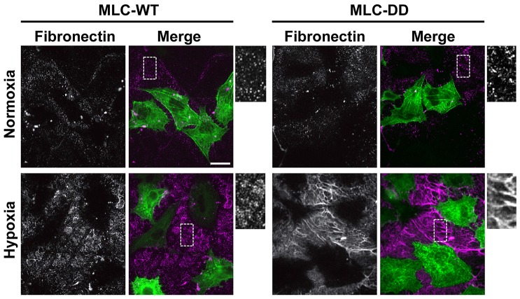
Fig. 3.
Phosphomimetic MLC rescues impaired fibrillogenesis with hypoxia. HK2 cells transiently expressing wild-type MLC–GFP (MLC-WT) or MLC-T18D/S19D–GFP (MLC-DD) were untreated (normoxia controls) or exposed to 1% hypoxia for 48 h. Representative images of cells immunolabeled for fibronectin. Scale bar: 20 µm.

