Fig. 1.
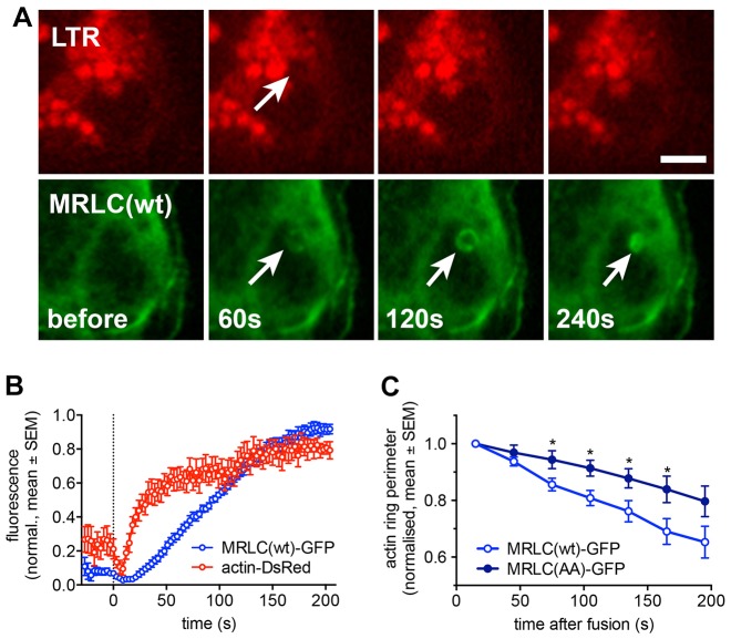
Myosin II is recruited to fused lamellar bodies following actin coat formation. (A) Simultaneous imaging of LTR (red) and MRLC–GFP (green) revealed recruitment of MRLC to lamellar bodies upon fusion with the plasma membrane. Lamellar body fusion with the plasma membrane is indicated by the selective decrease in LTR fluorescence due to diffusion of the LTR from the vesicle lumen (arrow, upper row). Time indicates time after fusion. Scale bar: 5 µm. (B) Time course of actin–dsRed (red) and MRLC(wt)–GFP (blue) fluorescence analysed in a circular region of interest around fusing lamellar body. Dashed line denotes time of fusion. Data represent mean±s.e.m. from eight individual fusions. (C) Expression of the non-phosphorylated MRLC mimic [MRLC(AA)–GFP] slowed down actin coat contraction significantly compared to expression of MRLC(wt)–GFP (*P<0.05 for 75–165 s; n = 15 and 11 for wt and AA, respectively).

