Fig. 2.
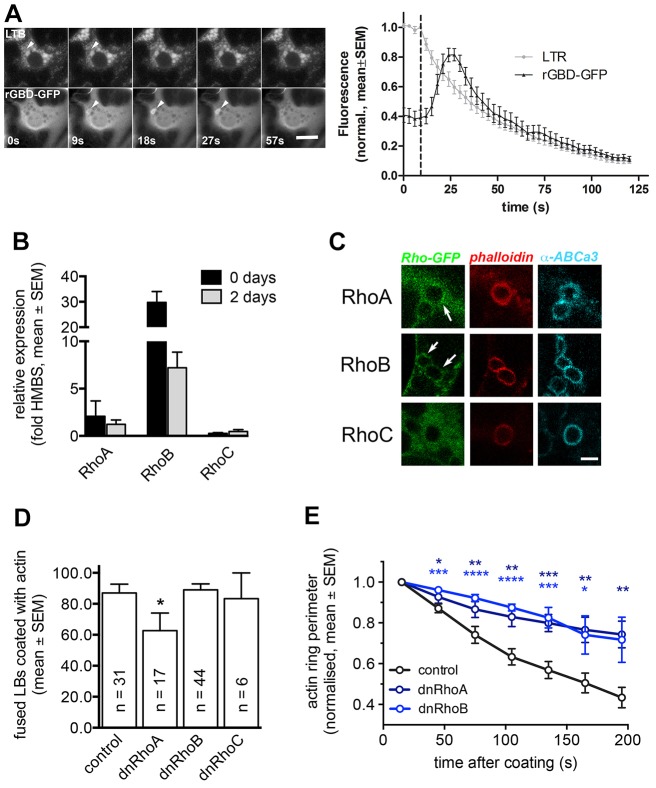
RhoA and RhoB translocate to fused lamellar bodies and probably regulate coat formation and contraction. (A) Left: The marker for active Rho GTPases, rGBD–GFP, transiently translocated to the fusing lamellar body (arrowhead, bottom row). Time of fusion was detected by LysoTracker fluorescence decrease (arrowhead, upper row). Scale bar: 10 µm. Right: LysoTracker fluorescence change (indicating vesicle fusion) and rGBD–GFP fluorescence change were measured in a circular region of interest around the fusing lamellar body (n = 17). (B) Real-time RT-PCR analysis of RhoA, RhoB and RhoC transcripts in freshly isolated rat ATII cells and ATII cells kept in culture for 2 days. Data are expressed as fold expression compared to the housekeeping gene Hmbs. Values are means from three individual cell isolations and are represented as mean±s.e.m. (C) Expression of Rho GTPase isoforms tagged with GFP revealed that only RhoA and RhoB (arrows) but not RhoC translocated to fused lamellar bodies following fusion with the plasma membrane. Fused lamellar bodies can be identified by the actin coat (phalloidin) on the lamellar body membrane (ABCa3). Scale bar: 2 µm. (D) Actin coating of fused lamellar bodies was significantly reduced in cells expressing dnRhoA-IRES–YFP, but not in cells expressing dnRhoB-IRES–YFP or dnRhoC-IRES–YFP. n represents number of cells analysed for each condition. Data are represented as means±s.e.m. (E) Expression of dnRhoA-IRES–YFP or dnRhoB-IRES–YFP in cells transfected with actin–dsRED resulted in a significantly decreased coat contraction compared to control cells (n = 19, 7 and 7 fusions for control, dnRhoA-IRES–YFP and dnRhoB-IRES–YFP, respectively). *P<0.05; **P<0.01; ***P<0.001; ****P<0.0001.

