Fig. 1.
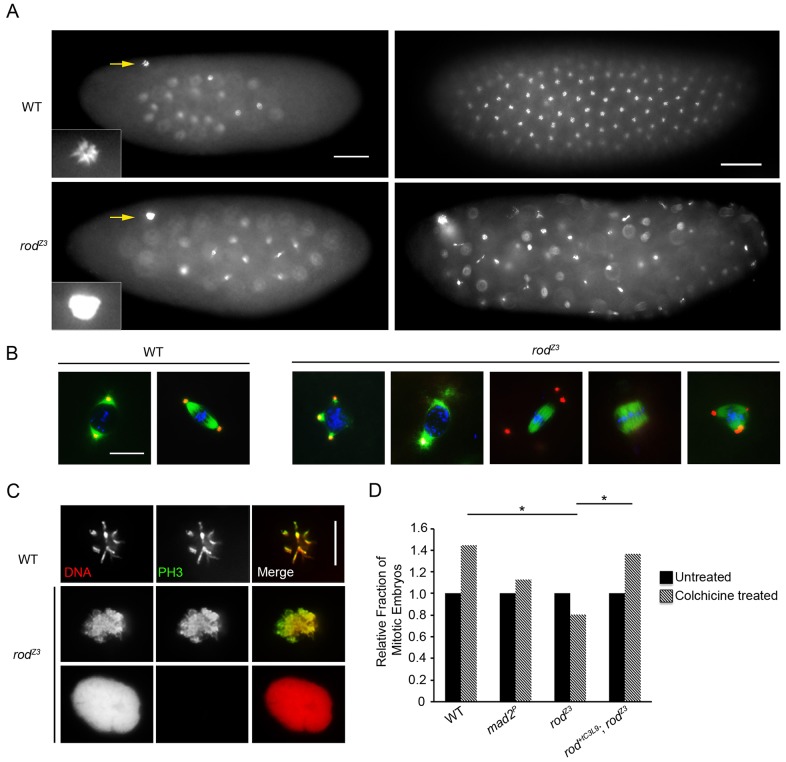
Embryos laid by rodZ3 mothers are defective in syncytial mitosis, polar body maintenance and the SAC. (A) Representative images of DNA-stained early (left) and late (right) syncytial embryos from wild-type (WT) and rodZ3 females. rodZ3-derived embryos contain large decondensed polar bodies (left). Also note the asynchrony of mitotic stages and the irregular distribution of nuclei in the rodZ3-derived embryos (right). Scale bars: 50 µm. Insets, larger images of polar bodies (marked by arrows). (B) Representative images of prophase and metaphase figures from WT and rodZ3-derived syncytial embryos stained for tubulin (green), centrosomin (red) and DNA (blue). rodZ3 mitotic figures have abnormal centrosome attachments. Broad acentrosomal and multipolar spindles are common. Scale bar: 10 µm. (C) Details of polar bodies from WT and rodZ3-derived syncytial embryos stained for PH3 and DNA. rodZ3 polar bodies are either PH3-positive (partially decondensed) or PH3-negative (fully decondensed). Scale bar: 10 µm. (D) The SAC is not functional in rodZ3-derived embryos. Relative change in the fraction of mitotic syncytial embryos after 30 min incubation in colchicine (compared with untreated, set at 1) for the indicated genotypes. rodZ3-derived embryos, like those derived from Mad2 null mothers (Mad2P, negative control), show no significant increase after treatment. *P<0.005; **P<0.0001 (Fisher's exact test; n = 80 embryos, except rescue-derived embryos where n = 140).

