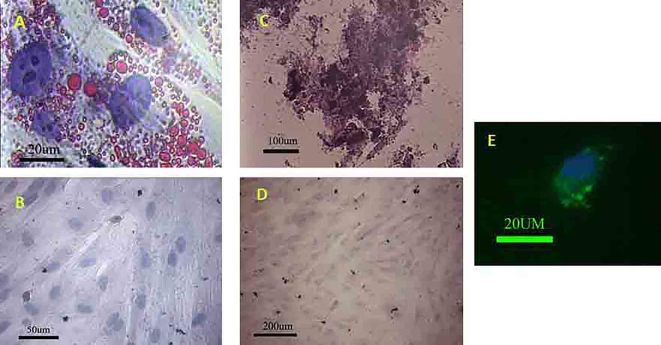
Figure 2.
(A) The MSCs could differentiate into adipocyte after exposing to the adipogenic media. (B) Negative control did not show any lipid droplet; oil red staining. The MSCs showed the potential to differentiate toward osteogenic lineage. (C) The red part contains calcium that reacted with alizarin red S. (D) Negative control did not show any Ca deposition; alizarin red S staining. MSCs premeabilized in the presence of FITC-dextran. (E) The green granule indicated the FITC-dextran was internalized by MSC.

