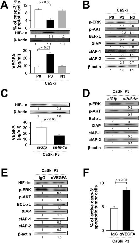
Figure 5. HIF-1α induces an anti-apoptotic program through the AKT and ERK pathways.
(A) Western blot analysis of a panel of pro- or anti-apoptotic factors in post-selection tumor cells (P3) transfected with siRNA against GFP or HIF-1α, or in pre-selection tumor cells (P0) transduced with empty vector or HIF-1α (P0/HIF-1α). β-actin was included as an internal control. (B) Western blot analysis of AKT, ERK, and p38 phosphorylation in P3 and P0 cells with indicated HIF-1α expression. (C) Western blot analysis of anti-apoptotic molecules in P0/HIF-1α cells treated with LY-294002 (LY; AKT inhibitor), PD-98059 (PD; ERK inhibitor), SB-203580 (SB; p38 inhibitor), or DMSO control (Con). (D) P0/HIF-1α cells treated as indicated were mixed with E7-specific CTLs. The frequency of apoptotic cells was determined by flow cytometry analysis of caspase-3 activation.

