
Figure 2. ABT induces CSF-1 expression, which promotes macrophage function.
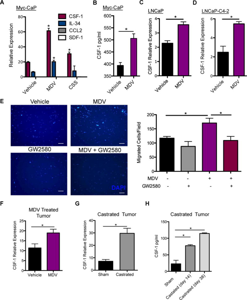
A) Quantitative RT-PCR analysis of TAM recruiting cytokines expression in Myc-CaP cells after 48 hrs of MDV3100 (10 μM) treatment. B) CSF-1 protein expression in Myc-CaP lysates after MDV3100 treatment. Quantitative RT-PCR of CSF-1 expression after 48 hrs of MDV treatment of C) LNCaP D) LNCaP-C4-2. (n = 3). E) 6 hr migration assay using RAW264.7 murine macrophages stimulated by conditioned media from Myc-CaP cells treated with MDV3100 or vehicle (DMSO), without or with the addition of 1 nM GW2580. DAPI staining of migrated RAW cells (left), migrated cell quantification of 10 fields/well at 4× magnification. Scaled bars represent 100 μm. (n = 3–6 per group). F) CSF-1 expression by RT-PCR from vehicle and MDV- treated Myc-CaP tumors. Tumor bearing mice were treated by daily oral gavage with vehicle, or MDV3100 (10 mg/kg) for 9 days. G) CSF-1 expression in Myc-CaP tumors by RT-PCR from sham (day 14 after sham surgery) and castrated (day 36 post castration) tumor bearing mice. H) Sera CSF-1 protein level analyzed by ELISA from sham and castrated mice at sham (day 14 after sham surgery), midpoint (day 14 after castration) and end point (day 38 after castration) was shown. *P < 0.05.