
Figure 4. CSF1R blockade effectively lowered tumoral and systemic macrophage levels.
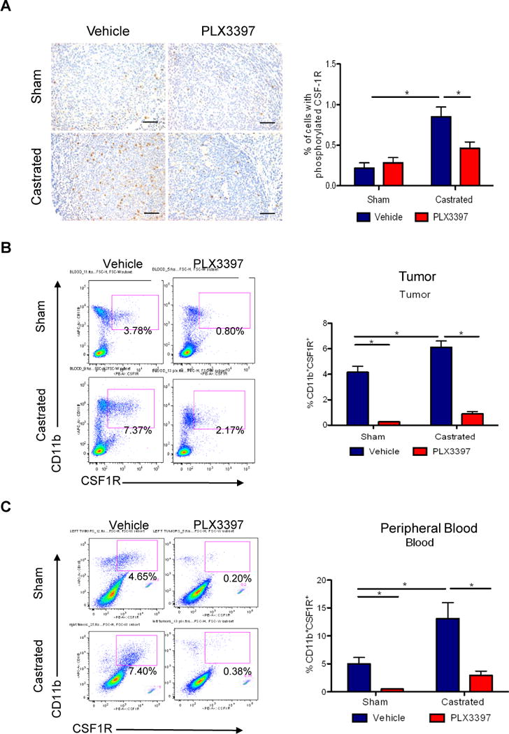
Myc-CaP tumor bearing mice received surgical castration or sham surgery when tumors reached 300–500 mm3, and then fed with control or PLX3397 chow daily for 36 days. A) Representative images of tumor sections stained (left panel) with anti-phosphorylated CSF1R (CSF1R-Tyr723) from 4 treatment groups. Scaled bars represent 100 μm. The right graph shows the quantification of IHC staining. B) Representative flow cytometry plots of B) total CD11B+CSF1R+ TAMs (left) in tumors, and C) total CD11B+CSF1R+ myeloid cells in peripheral blood, along with quantification of each (right). *P < 0.05. (n = 6–10 per group).