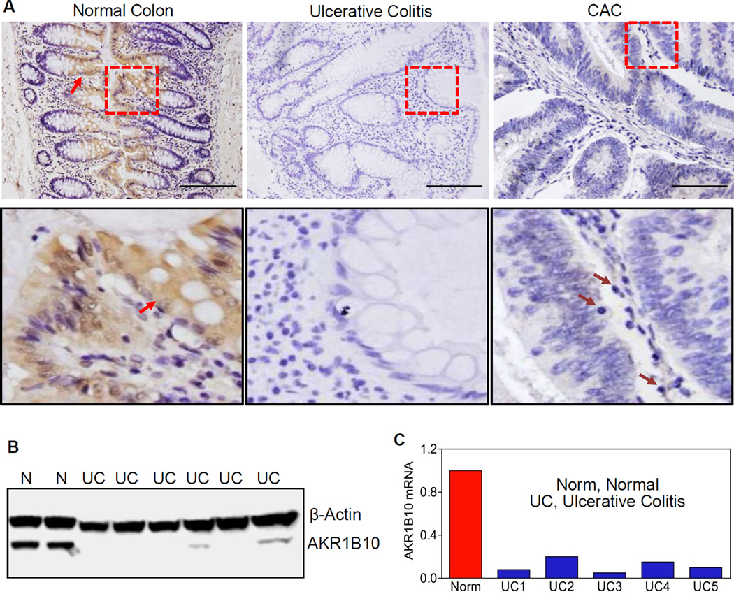
Figure 1. AKR1B10 expression in normal, ulcerative colitis and cancerous colon tissues.
(A) Immunohistochemistry in paraffin-embedded sections of normal colon, ulcerative colitis and associated tumor tissues. AKR1B10 protein was detected in normal colonic epithelium (arrows), but not in ulcerative colitis and tumor tissues. Lower panel: amplification of the squared areas. Purple arrows indicate infiltrated mononuclear cells in CAC. Scale bar, 100µm. (B) Western blot. N, normal colon tissues; UC, ulcerative colitis. (C) Real-time RT-PCR. The AKR1B10 mRNA level in normal colon is set up at 1.0, and AKR1B10 mRNA levels in ulcerative colitis tissues are presented as fold over normal. GAPDH mRNA was detected as an internal control.

