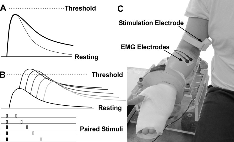
Fig. 1.
Hypothesis, protocol, and experimental setup. A: sketch of excitatory postsynaptic potential (EPSP) time course. The EPSP (thick line) in the hyperexcitable motoneuron has a slower rate of decay than the normal motoneuron (thin line). B: subthreshold double-stimulation protocol. C: experimental setup.

