Fig. 2.
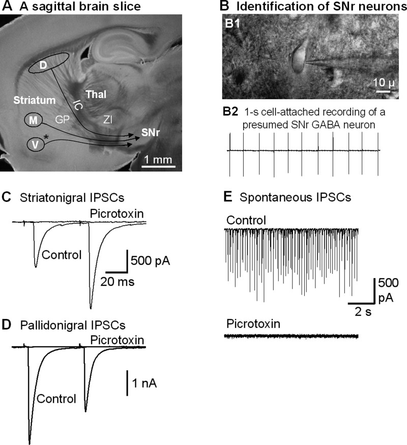
Focal stimulation in the striatum evokes striatonigral inhibitory postsynaptic currents (IPSCs) in SNr GABA neurons. A: picture of a live, 15° angular sagittal brain slice taken with a ×1 objective. The SNr and the striatal subregions are clearly identifiable. D, the stimulating site in the dorsal striatum; M, the stimulating site in the lower middle striatum; V, the stimulating site in the ventral striatum. GP, globus pallidus; IC, internal capsule; Thal, thalamus; ZI, zona incerta. *AC. B, B1: picture of a live brain slice, taken under a ×60 objective, shows a typical SNr neuron being patched. B2: typical spontaneous spikes (action potentials, ∼10 Hz) in a presumed SNr GABA neuron recorded in cell-attached mode. C: two pulses with a 50-ms interval in the dorsal striatum stimulation evoked striatonigral IPSCs in an SNr neuron that were blocked by 100 μM picrotoxin. Note the typical paired pulse facilitation in this example. D: an example of typical, depressing pallidonigral IPSCs evoked by 2 paired pulses. E: spontaneous IPSCs recorded in the absence of tetrodotoxin in an SNr GABA neuron were blocked by 100 μM picrotoxin.

