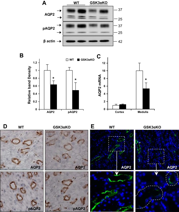
Fig. 3.
AQP2 abundance is reduced in GSK3αKO mice. Renal medullary AQP2 and phosphorylated (p)AQP2 (pSer256 AQP2) levels were reduced in GSK3αKO mice, as demonstrated by Western blot analysis (A) and quantitation of band density (B). C: AQP2 mRNA relative to β-actin was reduced in GSK3αKO mice. n = 6 mice/group. D: immunostaining showing reduced AQP2 and pAQP2 staining in the renal papilla of GSK3αKO mice. E: in the cortico-medullary junction of GSK3αKO kidneys, many collecting ducts showed very limited AQP2 (green; inset). Collecting ducts with normal AQP2 expression could also be seen in the same area (*). Magnification: ×63. *P < 0.05 compared with WT mice.

