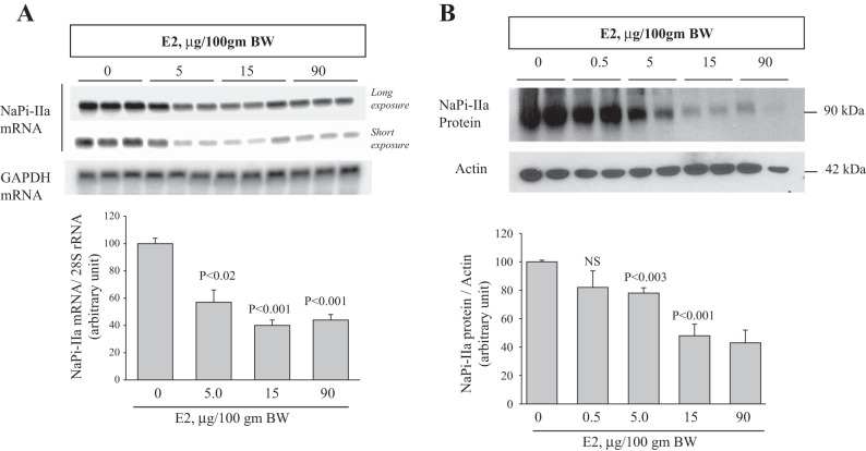
Fig. 2.
Dose-response effect of estrogen on NaPi-IIa expression in the kidney. A, top: representative Northern blots showing long and short exposure of NaPi-IIa mRNA (light bands) and GAPDH mRNA (dark band) in the renal cortex of rats injected daily for 3 days with indicated doses of estrogen vs. vehicle (0). GAPDH was used as a constitutive gene for the control of the equity of RNA loading into Northern gels. Bottom: corresponding densitometric analysis showing the mean of NaPi-IIa mRNA-to-GAPDH mRNA ratio (n = 4 rat in each group). B, top: representative immunoblots showing the abundance of NaPi-IIa and actin proteins in membrane fractions isolated from renal cortex of vehicle- or estrogen-treated rats. B, bottom: average densitometric analysis of NaPi-IIa/actin bands (n = 4 rat in each group). Each lane was loaded with 30 μg of total RNA or 40 μg of membrane proteins from a different rat. n = 4 rats in each group.

