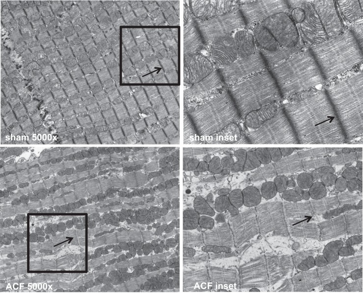
Fig. 6.
Eight-week ACF rats have disorganized cytoskeletal elements and loss of mitochondrial registry. Transmission electron microscopy (TEM) of the left ventricular (LV) myocardium in sham (top) and 8-wk ACF (bottom) hearts demonstrated pathological changes in mitochondrial morphology in ACF hearts compared with sham hearts. ACF mitochondria exhibited a loss of linear registry, clustering, disassociation with sarcomeres, and decreased electron density in ACF. The ACF myocardium also showed a breakdown in myofibrils, a decrease in Z-line electron density, and large gaps between sarcomeric units. Arrow in the top left image indicates normal Z-line electron density, whereas arrow in the bottom left image indicate the loss of electron density at the Z-lines due to ACF. Insets in the top left and bottom left images are expanded in the top right and bottom right images, respectively.

