Fig. 5.
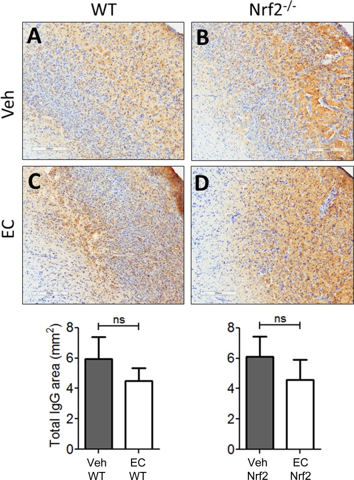
EC does not alter gross vascular permeability or hemorrhage frequency. Representative micrographs show mouse IgG immunohistochemical staining in sections counterstained with Cresyl violet. A and B: sections from vehicle-treated WT and Nrf2−/− mice showed expansive regions of mouse IgG staining that is indicative of blood brain barrier permeability, with no apparent difference in the expanse of parenchymal IgG immunoreactivity between genotypes. C and D: IgG immunoreactivity was abundant throughout the ischemic cortex in sections from EC-treated mice and appeared more prominent in Nrf2−/− mice relative to WTs. Bottom: quantification revealed no apparent difference in IgG immunoreactivity between vehicle- and EC-treated WT (P = 0.28) or Nrf2−/− mice (P = 0.92). Data are expressed as means ± SE. ns, not significant. Scale bars = 200 μm.

