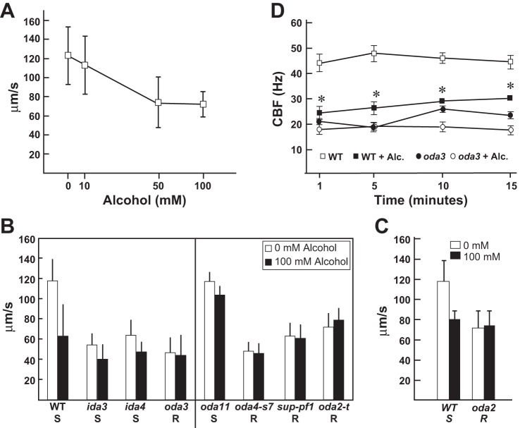
Fig. 1.
Alcohol treatment reduces ciliary beat frequency (CBF) and alters outer dynein arm (ODA) function via the β- and γ-heavy chain (HC). A: wild-type (WT) cells were treated with increasing levels of alcohol. Swimming speeds were measured and show a dose-dependent decrease in response to increasing alcohol concentration. B: motility analyses of WT cells and mutants strains defective in the inner dynein arm (IDA) and ODA motors (left) show that alcohol significantly reduces swimming velocities in WT cells and in mutant strains defective in the IDA motors [dyneins f (ida3) and dyneins a, b, and c (ida4)]. In contrast, the alcohol-induced decrease in swimming speed is not observed in oda3 (lacking the ODA). Motility analyses of oda HC mutants (right) show that alcohol reduces forward swimming speeds in oda11 (lacking the α-HC). In contrast, alcohol shows no ciliary-slowing effect in oda4-s7 (lacks β-HC motor domain), sup-pf1 [lacking 7 amino acids in the coiled-coil 1 (CC1) region of the dynein motor stalk domain], and oda2-t (lacks γ-HC motor domain). Thus WT, ida mutants, and oda11 are susceptible (S) to alcohol-induced ciliary dysfunction (AICD), whereas oda3 and the β- and γ-HC mutants are alcohol resistant (R). C: AICD is recapitulated in WT cell models, where the ciliary membrane is removed. Thus AICD is mediated by mechanisms intrinsic to the ciliary axoneme. Importantly, oda2 cell models are resistant to AICD and confirm that AICD targets the ODA motors. D: in the presence of 10 mM alcohol, CBF is significantly reduced in WT cells over the course of 15 min (*P ≤ 0.0001). CBF is not reduced by alcohol in a mutant that lacks the ODA (oda3). Measurements were performed using Sisson-Ammons Video Analysis (42).

