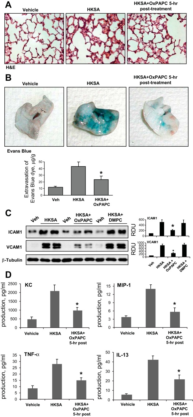
Fig. 7.
Effects of OxPAPC posttreatment on HKSA-induced tissue injury Evans blue extravasation and inflammatory markers. C57BL/6J mice were challenged with vehicle or HKSA (2 × 10⁸ cells/mouse it) with or without OxPAPC posttreatment (1.5 mg/kg, 5 h after HKSA). Analysis of lung injury was performed 48 h after HKSA challenge. A: histological analysis of lung tissue by hematoxylin and eosin staining (×40 magnification). B: Evans blue dye (30 ml/kg iv) was injected 2 h before termination of the experiment. Lung vascular permeability was assessed by Evans blue accumulation in the lung tissue. The quantitative analysis of Evans blue-labeled albumin extravasation was performed by spectrophotometric analysis of Evans blue extracted from the lung tissue samples; *P < 0.05 vs. HKSA alone; n = 3. C: ICAM1 and VCAM1 protein expression in lung tissue samples was evaluated by immunoblotting analysis. Membrane probing with β-tubulin antibody was used as a normalization control. The results of Western blot quantitative densitometry are presented as means ± SD; n = 4. D: levels of mouse cytokines KC, MIP-1, TNF-α, and IL-13 were measured in BALF samples by ELISA assay. Data are expressed as means ± SD of 4 independent experiments; *P < 0.05.

