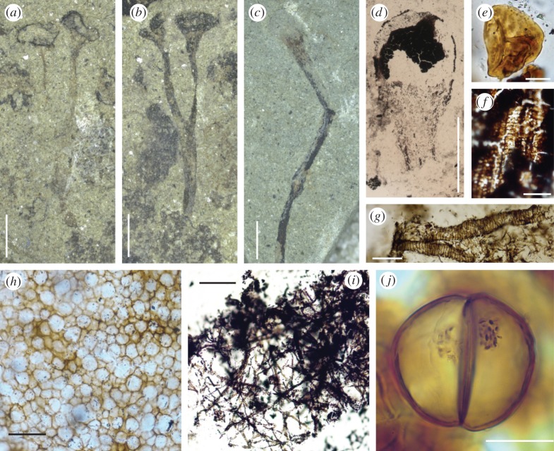
Figure 2.
New light micrographs of material illustrated by Lang [1]. Original number and figure numbers in brackets. (a) Cooksonia pertoni. Lectotype. Přídolí. V58011. (124 Z b; Plate 8, fig. 8). Coalified material has been removed, possibly using cellulose acetate, for further analysis. Scale bar, 2 mm. (b) Counterpart of (a). V58010. Black patches possibly of Nematothallus. Scale bar, 2 mm. (c) Cooksonia hemisphaerica. Lochkovian. V58022. (186; Plate 9, fig. 33). Scale bar, 2 mm. (d) C. hemisphaerica sporangium recovered using cellulose acetate. V54592. (1062; Plate 9, fig. 34). Scale bar, 1 mm. (e) Spore recovered from a sporangium of C. pertoni. V54995. (1105: Plate 8, fig. 11). Scale bar, 10 μm. (f) Tracheids recovered using cellulose acetate from a sterile axis, thought by Lang to belong to C. hemisphaerica. Lochkovian. V55040. (1150; Plate 9, fig. 36). Scale bar, 50 μm. (g) Banded tubes on cellulose acetate sheet. Přídolí. (962; Plate 11, fig. 60). Scale bar, 50 μm. (h) Nematothallus ‘cuticle′ on a cellulose acetate sheet. Přídolí. V54697. Scale bar, 50 μm. (i) Typical wefts of tubes assigned to Nematothallus. V54851. (Not figured, but cf. plate 11). Scale bar, 160 μm. (j) Dyad in elongate cylindrical spore mass. Přídolí. V54654. (764; Plate 13, fig. 109). Granular material probably represents condensed cell contents. Scale bar, 20 μm.

