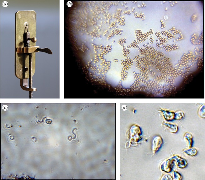
Figure 4.
(a) Replica of a single-lens microscope by Leeuwenhoek (Image by Jeroen Rouwkema. Licensed under CC BY-SA 3.0 via Wikimedia Commons). (b,d) Photomicrographs taken using simple single-lens microscopes including one of Leeuwenhoek's originals in Utrecht, by Brian Ford (Copyright © Brian J. Ford). (b) An air-dried smear of Ford's own blood through the original van Leeuwenhoek microscope at Utrecht, showing red blood cells and a granulocyte with its lobed nucleus (upper right; about 2 µm in diameter). (c) Spiral bacteria (Spirillum volutans) imaged through a replica microscope with a lens ground from spinel; each bacterial cell is about 20 µm in length. (d) The intestinal protist parasite Giardia intestinalis imaged through a replica soda-glass produced by Brian Ford [28,29].

