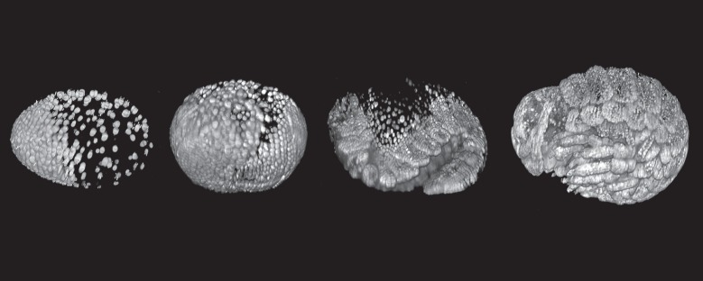
Figure 1.

Following crustacean development in a live embryo at single-cell resolution with modern techniques. The figure shows four time points during the development of an embryo of the amphipod crustacean Parhyale hawaiensis, imaged using multi-view fluorescence light-sheet microscopy (lateral views, anterior to the left). The nuclei are fluorescently labelled using a transgenic construct. The image at the left shows an early stage while cleavage nuclei are aggregating to form the embryonic primordium; the image at the right shows a late differentiation stage, with the forming antennae and limbs clearly visible. A full movie of Parhyale development is available at the link http://www.cell.com/pictureshow/lightsheet2, which also explains further how the data were collected. Images courtesy of Anastasios Pavlopoulos.
