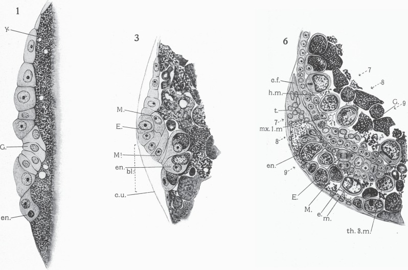
Figure 2.
Sections of fixed and stained embryos of Hemimysis, as figured in the plates accompanying Manton's paper [1], showing her attribution of cell identities. Plate 1: The beginning of gastrulation. A single layer of cells overlies the yolk (y). The future germ line cells (G) are starting to buckle inwards at the blastopore. Plate 3: A later stage in gastrulation, with ectodermal teloblast (E), mesodermal teloblast (M) and endoderm (en) all identified in the region of the blastopore (bl). Plate 6: A later stage, after the caudal end (c.f.) of the embryo has flexed forward, showing the rich detail of cell differentiation in these preparations. Copyright © The Royal Society.

