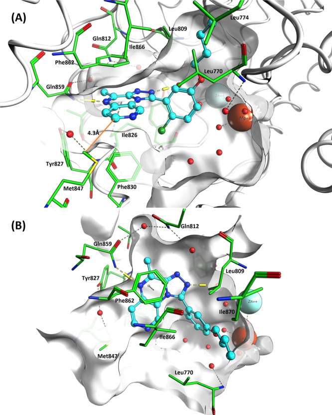
Figure 1.

Molecule 9 (cyan color) docked into PDE2A crystal structure solved with 3 (PDB 4D08). Viewed from the entrance to the active site (A) and top down (B). Important amino acids are highlighted in green, and catalytic Zn2+ and Mg2+ ions are shown, along with active site water molecules (red spheres). Distance between 9 and Met847 highlighted in orange.
