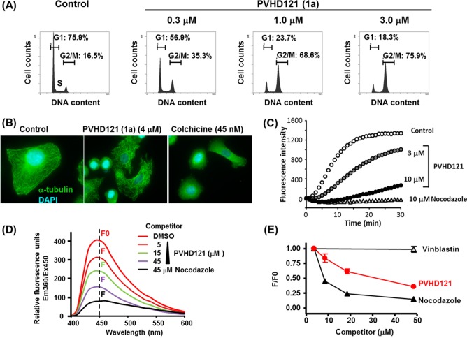
Figure 1.
(A) Flow cytometric analysis of cell cycle distribution in A549 cells. (B) Immunomicroscopic analysis of tubulin destabilization in A549 cells. (C) In vitro tubulin polymerization assay. (D) Competitive displacement binding assay on the colchicine-binding site of tubulin. (E) Concentration-dependent binding of 1a to the colchicine-binding site.

