Fig. 6.
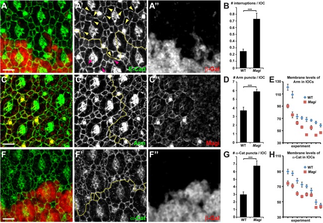
Magi mutant has abnormal AJs. (A-A″,C-C″,F-F″) 24-h APF pupal eye discs with Magi mutant clones marked by the absence of β-galactosidase (red, A″,F″) or the absence of Magi (red, C″). Yellow lines (A′,C′,F′) mark the boundaries between wild-type and mutant tissues. (A) In Magi mutant tissue, E-Cad staining (green, A′) is frequently interrupted (yellow arrowheads) compared with wild type (purple arrowheads). (C,F) In Magi mutant tissue, β-catenin (Arm; green, C′) and α-catenin (α-Cat; green, F′) are less cortical and accumulate in cytoplasmic vesicles in IOCs. (B,D,G) Quantification of E-Cad interruptions (B), Arm vesicles (D) and α-Cat vesicles (G) in wild-type and Magi mutant tissues corresponding to A, C and F, respectively. s.e.m. is shown; ***P<0.001 (unpaired t-test). (E,H) Quantification of Arm (E) and α-Cat (H) membrane levels between IOCs in wild-type and Magi mutant tissues corresponding to C and F, respectively. Eight independent pairs of wild-type and Magi mutant tissues are shown and plotted by decreasing arbitrary average pixel intensity. Levels in wild type are always higher than those in Magi. s.e.m. is shown; P<0.001 (paired t-test). Scale bars: 5 µm.

