Fig. 6.

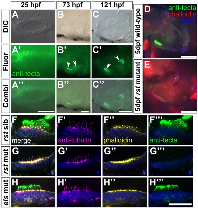
Expression of α-Tectorin protein in wild-type and rst mutant embryos. (A-C″) Immunofluorescence analysis showing that α-Tectorin protein is localised to the anterior OV at 1 dpf and to the two otolithic membranes at 3 and 5 dpf (arrowheads). Anterior is to the left. (D) Confocal image showing α-Tectorin protein localisation to the utricular otolithic membrane and to cells in the utricular epithelium (green) in a 5 dpf wild-type (AB strain) embryo. Anterior left, dorsal up. (E) In a 5 dpf rst embryo, α-Tectorin is not localised to the otolithic membrane of the utricular macula, although there is a low level of protein detectable within the utricular epithelium. (F-H‴) Confocal images of utricular maculae from 5 dpf embryos; lateral views, anterior to left. Nuclei are stained with DAPI (blue), hair cells and kinocilia with anti-acetylated Tubulin antibody (magenta), filamentous actin and hair cell stereociliary bundles with Alexa647-phalloidin (yellow), and α-Tectorin by antibody (green). (F-F‴) Phenotypically wild-type 5 dpf rst sibling embryo, showing strong staining for α-Tectorin in the utricular otolithic membrane, and protrusion of the hair cell kinocilia into this membrane. α-Tectorin staining is also visible in the saccular otolithic membrane (asterisk, F). (G-G‴) 5 dpf rst mutant embryo with no extracellular α-Tectorin stain. There is a weak α-Tectorin signal at the apical surface of the hair cells (G‴). Kinocilia appear normal (G′). (H-H‴) 5 dpf eis mutant embryo with normal expression and localisation of α-Tectorin and normal kinocilia. Scale bars: 50 µm in A-B″; 100 µm in C-E; 20 µm in F-H‴.
