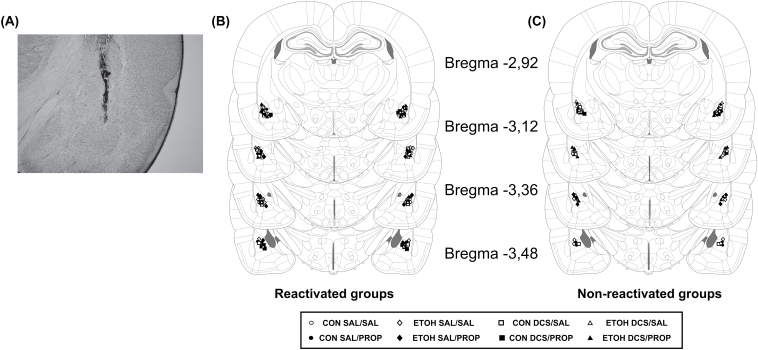
Figure 6.
Placement of infusion cannulas.
(A) Photomicrograph of a coronal brain section showing the location of the infusion site in the BLA. Magnification: 25x. Schematic representation of coronal sections of the rat brain showing the cannula tip placements for reactivated groups (B) and non-reactivated groups (C) in the BLA for Experiment 3 (adapted from Paxinos and Watson, 2009). BLA, basolateral amygdala; CON, control rats; DCS, d-cycloserine; ETOH, ethanol-withdrawn rats; PROP, propranolol; SAL, sterile saline.

