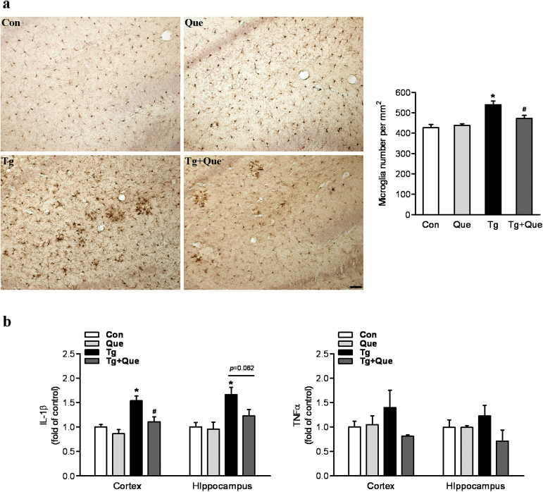
Figure 3.
Quetiapine attenuates microglial activation and reduces proinflammatory cytokines in APP/PS1 mice. a, Representative immunohistochemical staining with anti-ionized calcium binding adapter molecule 1 (Iba1) in hippocampus following the treatment. The scale bar represents 50 μm. Quantification of the number of Iba positive cells was shown in the graph. Two-way analysis of variance (ANOVA) showed microglial cell density was increased in transgenice mice and decreased following quetiapine treatment. (b) Enzyme-linked immunosorbent assay (ELISA) analysis of selected proinflammatory cytokines. Two-way ANOVA showed quetiapine treatment greatly attenuated the increase of interleukin 1β (IL-1β) in the cortex of transgenic mice. No statistcial significance was detected in the level of tumor necrosis factor α (TNFα). Data are expressed as means±SEM, n=5 to 8 mice per group. * P<.05 vs Con; # P<.05 vs transgenic + water.

