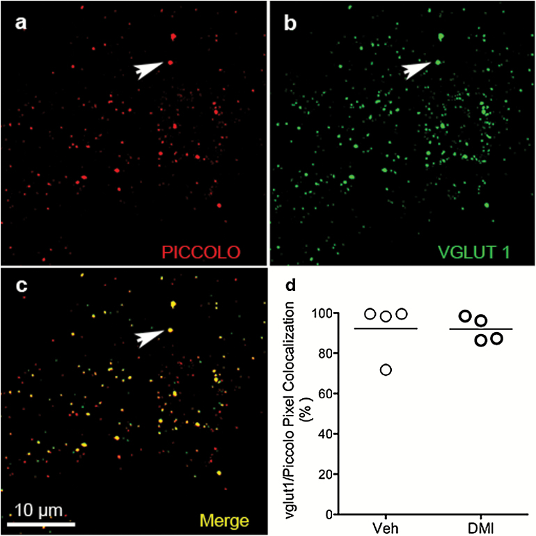
Figure 2.
Relative distribution of excitatory synaptic terminals within medial prefrontal cortex (mPFC). Dual staining for active-zone marker Piccolo (white arrow) (a) and transporter glutamate vesicular transport 1 protein (VGLUT1) (white arrow) (b) shows high degree of colocalization (white arrow) (c). Scale bar=10 μm. (d) Summary of quantitative data of colocalized punctate on dual-stained mPFC sections from rats treated with either vehicle (Veh) or chronic desipramine (DMI), obtained using VIS Image Analysis Software (Visiopharm, Hørsholm, Denmark). P value, ns. (N=4; n=5).

